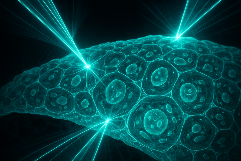
Image generated by AI
Imagine being able to watch a cancer cell divide, observe a neuron fire, or detect a viral infection spreading through tissue—all without injecting dyes, radioactive tracers, or genetically modifying the cells themselves. This is no longer science fiction. Scientists have developed a suite of optical and physical techniques that can image living cells and tissues in their natural state, revealing their secrets through light, sound, and electromagnetic fields alone. Yet most people remain unaware that such powerful, non-invasive windows into the microscopic world even exist.
The ability to see inside living tissue without adding foreign markers represents a fundamental shift in how we diagnose disease, conduct research, and understand the machinery of life. These marker-free imaging techniques bypass a major limitation of conventional methods: foreign dyes and tracers can alter cellular behavior, interfere with biological processes, and sometimes prove toxic to the very organisms we’re trying to study. In hospitals and laboratories around the world, researchers are racing to perfect these label-free approaches, driven by the promise of faster diagnoses, safer imaging for patients, and a deeper understanding of biological systems as they truly exist in nature.
What Is Cellular and Tissue Imaging Without Invasive Markers?
Cellular and tissue imaging without invasive markers refers to a family of techniques that visualize the structure, composition, and dynamics of living cells and tissues without introducing external labels, fluorescent dyes, or contrast agents. Instead, these methods exploit the intrinsic optical and physical properties of biological molecules—their ability to absorb, scatter, or refract light; to vibrate at specific frequencies; or to respond to electromagnetic fields. The term “non-invasive” emphasizes that the measurement process does not perturb the biological system being observed, allowing researchers to study cells in their native, unaltered state. The techniques range from optical methods like label-free microscopy and Raman spectroscopy to acoustic approaches like ultrasound biomicroscopy and mechanical techniques like atomic force microscopy.
The development of marker-free imaging accelerated significantly in the late 20th century, particularly after the invention of confocal microscopy in the 1980s and the subsequent refinement of techniques like coherent anti-Stokes Raman scattering (CARS) in the 1990s. Pioneers like Watt Webb and Tony Wilson at Cornell University developed confocal approaches, while researchers like Xiaolian Sunney Xie at Harvard extended these ideas into the realm of label-free vibrational imaging. Unlike fluorescence microscopy, which relies on added dyes that emit light when excited, these newer methods read the biological signatures already present in cells—the vibrations of chemical bonds, the refractive index differences between cellular structures, and the spatial organization of molecules themselves.
The Basic Principle
At the heart of label-free imaging lies a deceptively simple idea: matter interacts with light and other forms of energy in characteristic ways, and by carefully measuring these interactions, we can infer what’s there. Every molecule has a unique vibrational fingerprint—specific frequencies at which its chemical bonds oscillate. When infrared light or Raman laser light shines on a cell, certain wavelengths are absorbed or scattered by these vibrating bonds, creating a spectral signature as distinctive as a fingerprint. By scanning this spectrum across a sample and mapping which wavelengths are absorbed at each location, a microscope can build up an image of molecular composition with remarkable chemical specificity—and it does so without ever adding a label.
Think of it like identifying people in a crowded room by their voices alone. Each person’s voice has a unique frequency signature—their accent, pitch, and cadence. If you close your eyes and listen carefully, you can learn who’s talking without them wearing name tags or raising their hands. Similarly, label-free microscopy listens to the “voice” of molecules. When a laser beam hits cellular material, water molecules, lipids, and proteins all “sing” at different frequencies. A detector tuned to the right frequencies can distinguish one molecule from another based purely on its vibrational properties, creating a detailed chemical map of the tissue without any artificial markers.
Why It Matters in the Real World
The clinical implications of label-free imaging are profound and far-reaching. Fluorescent markers, while powerful, have significant drawbacks: they can be phototoxic, generate reactive oxygen species that damage cells, undergo photobleaching that degrades image quality, and may not perfectly represent native cellular conditions. For cancer diagnosis, label-free techniques can reveal the density and organization of collagen, lipids, and proteins that distinguish healthy tissue from malignant growths—changes that occur naturally and need no amplification through artificial dyes. In neuroscience, researchers use label-free imaging to study myelin, the insulating sheath around nerve fibers, revealing how diseases like multiple sclerosis progressively damage this structure. For drug development, pharmaceutical companies employ these techniques to observe how potential medications affect cell structure and metabolism in real-time, without the confounding effects of fluorescent labels.
Beyond the laboratory, label-free imaging is beginning to enter clinical practice. Hospitals use optical coherence tomography (OCT), a label-free technique that works much like ultrasound but uses infrared light instead of sound waves, to examine retinal disease, coronary plaques, and early skin cancers. Dermatologists deploy Raman spectroscopy to distinguish melanoma from benign moles by analyzing the chemical composition of suspicious lesions. Surgical teams employ two-photon excited fluorescence and harmonic generation microscopy—which use the intrinsic properties of tissue collagen to generate images without added dyes—to identify tumor margins and guide resection in real-time. These applications have moved from research curiosities to practical tools that improve patient outcomes.
Recent Breakthroughs in Cellular and Tissue Imaging Without Invasive Markers
The past two to three years have witnessed remarkable acceleration in this field. In 2023 and 2024, several teams reported significant advances in Raman spectroscopy and coherent Raman imaging, achieving unprecedented spatial resolution and speed. Researchers at MIT and collaborating institutions published work demonstrating that stimulated Raman scattering (SRS) microscopy can now image entire tissues sections in minutes rather than hours, with cellular-level resolution and without photobleaching—a dramatic improvement over fluorescence methods. Simultaneously, groups working on optical coherence tomography have achieved three-dimensional imaging of living organisms and tissues at depths previously unreachable, using adaptive optics and new wavelengths that penetrate deeper into tissue while maintaining sharp focus. Label-free techniques for imaging neural activity, based on intrinsic optical signals and voltage-sensitive dyes (which themselves count as non-invasive compared to calcium indicators), are now revealing brain circuits with unprecedented detail in awake, behaving animals.
The current frontier of research involves combining multiple label-free modalities into integrated platforms. Teams are developing microscopes that simultaneously acquire Raman spectra, harmonic generation images, and autofluorescence to create multi-dimensional chemical maps of tissue. Machine learning algorithms are being trained to interpret these complex datasets and recognize disease states or cellular conditions directly from the spectral signatures, without human interpretation. Other researchers are pushing toward real-time, in vivo imaging—watching cells and tissues in living organisms as biological processes unfold. Questions remain about how to penetrate even deeper into thick tissues, how to maintain sufficient resolution over large fields of view, and how to translate these tools into cost-effective diagnostic devices accessible in clinical settings worldwide.
Why Cellular and Tissue Imaging Without Invasive Markers Matters for the Future
As personalized medicine and precision diagnostics become central to healthcare, label-free imaging offers a pathway to faster, safer, and more informative patient evaluations. A surgeon performing tumor resection could use Raman spectroscopy or coherent imaging to assess margins in real-time, ensuring complete removal while preserving healthy tissue. A pathologist analyzing a biopsy could obtain full three-dimensional chemical information about cellular organization and composition within minutes, rather than days, accelerating diagnosis and treatment decisions. In drug discovery, pharmaceutical companies could screen thousands of compounds by directly observing their effects on cellular ultrastructure and metabolism, predicting efficacy and toxicity with greater accuracy. For rare diseases and pediatric medicine, where repeated imaging is often necessary and radiation exposure must be minimized, label-free optical techniques could become the imaging modality of choice.
Yet significant challenges remain. Label-free techniques generally require longer data acquisition times than fluorescence microscopy, making them less suitable for fast-moving phenomena or high-throughput screening applications. Penetration depth in thick tissues remains limited by light scattering, constraining the technique’s applicability to superficial structures or thin tissue slices—though optical clearing methods and new wavelengths are steadily improving this. The interpretation of spectral data demands sophisticated algorithms and often deep domain expertise, creating barriers to widespread adoption. Cost and accessibility remain concerns; while some label-free approaches are becoming more affordable, many still require expensive lasers and specialized expertise not yet available in most hospitals and clinics.
Key Takeaways
- Label-free cellular and tissue imaging visualizes living cells by detecting the intrinsic optical and physical properties of biological molecules, such as their vibrational frequencies and refractive indices, without adding external dyes or markers.
- The core mechanism relies on the characteristic “fingerprints” that molecules produce when interacting with light—especially infrared or Raman light—which vary by chemical composition and allow discrimination between cell types and disease states.
- The most promising clinical applications include real-time surgical guidance for tumor resection, rapid pathological diagnosis, and non-invasive assessment of retinal, coronary, and skin diseases, all with reduced phototoxicity compared to fluorescence-based approaches.
- Recent breakthroughs have dramatically improved the speed and resolution of techniques like stimulated Raman scattering and optical coherence tomography, while machine learning algorithms are beginning to automate interpretation of spectral data for clinical decision-making.
- As precision medicine and personalized diagnostics gain importance, label-free imaging represents the future of non-invasive, information-rich cellular imaging that could transform diagnosis and treatment while bypassing the limitations and risks of conventional contrast agents.
Explore TED Talks on Cellular and Tissue Imaging Without Invasive Markers:
TED content is used under CC BY-NC-ND 4.0. © TED Conferences, LLC.
Frequently Asked Questions
How do label-free imaging techniques detect cellular structures without fluorescent dyes or radioactive tracers?
Label-free techniques use intrinsic physical properties of cells and tissues—such as their natural optical properties, acoustic impedance, or electromagnetic interactions—to generate contrast without external markers. Methods like optical coherence tomography, Raman spectroscopy, and photoacoustic imaging exploit these inherent biological characteristics to visualize cellular components.
Why can injected dyes and radioactive markers interfere with the biological processes researchers are trying to study?
Foreign markers can alter cellular behavior by binding to molecules, changing protein function, triggering toxicity, or disrupting normal metabolic pathways, which means researchers may be observing artificially modified cells rather than cells in their natural state. This interference can lead to false conclusions about normal biological function.
What are the main physical mechanisms—such as light, sound, or electromagnetic fields—that enable marker-free cellular imaging?
Common mechanisms include optical techniques (light scattering and absorption), acoustic methods (ultrasound and photoacoustics), and electromagnetic approaches (electrical impedance and magnetic resonance). Each exploits different physical interactions between the imaging probe and the tissue's intrinsic molecular composition and structure.
Can label-free imaging techniques achieve the same cellular resolution and speed as conventional fluorescence imaging methods?
Label-free methods continue to improve in resolution and speed, with some techniques now approaching sub-micrometer resolution, though most remain slower than conventional fluorescence; however, they compensate by providing real-time, continuous monitoring of living cells without the temporal limitations of photobleaching or phototoxicity inherent to dye-based imaging.