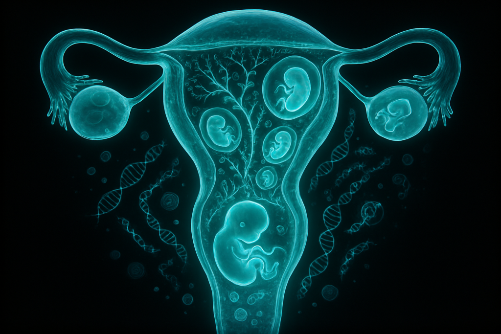
Image generated by AI
A fertilized egg becomes a thinking, breathing human being in just nine months—a transformation so profound that it staggers the imagination, yet so common that we rarely pause to marvel at its intricacy. From the moment sperm meets egg, an elaborate molecular choreography begins, orchestrating the creation of roughly 37 trillion cells, each finding its place in an increasingly complex architecture. Recent advances in imaging, genetics, and molecular biology have unveiled astonishing details about how this process unfolds, revealing that the fetus is not a passive passenger but an active participant in its own development, communicating with its mother through chemical signals and even influencing her immune system.
Understanding pregnancy and fetal development has never been more urgent or more consequential. Today, nearly 140 million babies are born annually worldwide, yet maternal and fetal complications remain a leading cause of disability and death in women of reproductive age, particularly in low-resource settings. Simultaneously, new technologies—from non-invasive prenatal testing to 3D ultrasound and artificial intelligence—are transforming our ability to monitor fetal health, diagnose abnormalities early, and intervene before birth. This convergence of biomedical innovation and clinical necessity makes fetal development one of the most dynamic frontiers in modern medicine, promising safer pregnancies and healthier outcomes for both mother and child.
What Is Pregnancy and Fetal Development?
Pregnancy is the biological process in which a fertilized ovum develops into a fetus and ultimately a newborn baby, typically lasting 40 weeks (280 days) from the last menstrual period. Fetal development refers specifically to the transformation of a single-celled embryo into a fully formed human being, beginning with implantation in the uterine wall around day six to twelve after fertilization and continuing through birth. The process is conventionally divided into three trimesters of roughly thirteen weeks each, each marked by distinct developmental milestones: the first trimester sees the formation of major organs and systems, the second trimester witnesses rapid growth and the development of sensory capabilities, and the third trimester emphasizes maturation and preparation for extrauterine life. Unlike organisms that reproduce asexually or in external environments, human fetal development is uniquely characterized by an intimate metabolic and immunological partnership between mother and fetus, mediated by the placenta—a temporary organ that is neither entirely fetal nor maternal but fundamentally chimeric.
The scientific understanding of human reproduction has evolved dramatically over centuries. In antiquity, figures like Hippocrates and Galen speculated about fetal development based on philosophical reasoning and dissection of non-human animals, often concluding that the fetus was a miniature human simply enlarging in size. During the Renaissance, anatomists like Andreas Vesalius and later William Harvey made detailed observations of pregnant organs, yet mysteries remained about fertilization itself. It wasn’t until the nineteenth century, with the advent of modern microscopy, that scientists like Karl Ernst von Baer identified the egg cell and Oscar Hertwig demonstrated that fertilization involved the fusion of sperm and egg nuclei. The twentieth century brought molecular biology’s revelations about genes and hormones, while the twenty-first century has exploded with epigenetic insights, revealing that genes are merely the starting script—their expression shaped profoundly by environmental, maternal, and developmental cues.
What the Research Shows
The developmental program unfolds through a series of precisely timed cellular processes that collectively transform a sphere of identical cells into the diverse tissues and organs of a human body. Within days of fertilization, the embryo undergoes cleavage—rapid cell divisions without growth—creating a ball of cells called the morula, which then cavitates to form the blastocyst, a structure containing two distinct cell populations: the inner cell mass (which gives rise to the fetus) and the trophectoderm (which forms the placenta and supporting tissues). Implantation, occurring around day six to twelve, is a delicate invasion of the uterine wall by trophoblastic cells, triggering a cascade of immunological tolerance that allows the genetically foreign fetus to be accepted by the maternal immune system—a feat so remarkable that immunologist Peter Medawar famously called the fetus “the most successful parasite.” Following implantation, gastrulation reorganizes the embryonic cells into three primary germ layers (ectoderm, mesoderm, and endoderm), each of which generates specific tissues: the ectoderm becomes the nervous system and skin, the mesoderm becomes muscle and bone, and the endoderm becomes the digestive and respiratory systems.
Think of early development like a city being built from blueprint to skyline. The genetic blueprint arrives at conception as DNA from both parents, but the “construction regulations” that determine how that blueprint is interpreted emerge from maternal hormones, placental signals, and even the physical environment of the uterus itself. If you magnified a developing embryo to the size of a city block, you would see waves of cells migrating like commuters heading to work, some traveling hundreds of micrometers to reach their destinations—distances that, when scaled up, would be cross-country journeys. The notochord, a transient structure running down the embryo’s midline, acts like a master-control tower, releasing chemical signals called morphogens that diffuse through neighboring tissues, specifying their fate: cells close to high concentrations of a particular morphogen become one structure, while those farther away become another. This elegant system of positional information, discovered by pioneers like Lewis Wolpert, allows a relatively simple set of molecular signals to generate breathtaking complexity.
What This Means for Patients and Science
Understanding the mechanisms of fetal development has profound implications for preventing birth defects and managing complications during pregnancy. Approximately 3 to 5 percent of babies are born with some form of congenital abnormality, ranging from minor to life-threatening, and many result from disruptions during critical windows of development when specific organs are being formed—a concept called teratogenic sensitivity. Maternal infections (such as Zika virus or rubella), certain medications (including some anti-seizure drugs and retinoids), nutritional deficiencies (particularly folate), maternal metabolic disorders (diabetes, thyroid disease), and environmental exposures (alcohol, heavy metals) can all interfere with normal developmental processes, sometimes with devastating consequences. By understanding what normally controls development, researchers can identify vulnerable stages, screen for risk factors, and design interventions to prevent or mitigate harm. Women taking folic acid supplements before and during early pregnancy, for instance, significantly reduce the risk of neural tube defects—a direct application of developmental biology knowledge.
In clinical practice today, this knowledge manifests through several technologies and approaches. Ultrasound screening at 11 to 14 weeks, measuring the nuchal translucency (fluid behind the fetal neck), combined with maternal blood tests for pregnancy-associated proteins, identifies pregnancies at higher risk for chromosomal abnormalities like Down syndrome without the miscarriage risk of invasive amniocentesis. Non-invasive prenatal testing (NIPT), which analyzes fetal DNA fragments circulating in maternal blood, offers earlier and more accurate detection of genetic abnormalities. Three-dimensional and four-dimensional ultrasound imaging now allows visualization of fetal anatomy with stunning detail, enabling diagnosis of structural anomalies before birth and, in some cases, planning for postnatal intervention or specialized delivery procedures. Fetal echocardiography can detect congenital heart defects in utero, allowing cardiologists to prepare for surgery immediately after birth. Maternal-fetal medicine specialists use this developmental knowledge to time interventions appropriately, understanding that some conditions are best addressed prenatally, while others benefit from allowing development to progress further before intervention.
Recent Breakthroughs in Pregnancy and Fetal Development
The past three years have witnessed remarkable advances in our ability to visualize and manipulate early development. In 2021 and 2022, researchers led by teams at the Weizmann Institute and Cambridge University created synthetic embryos from mouse embryonic stem cells, developing to mid-gestation stages without a sperm or egg—demonstrating that the complex self-organization properties of embryonic tissues could generate recognizable body plans from cellular “ingredients” alone. While these synthetic embryos remained incomplete and the technology remains distant from human application, they offer unprecedented insights into the self-organizing principles underlying development. Simultaneously, advances in single-cell sequencing have allowed scientists to catalog every cell type in developing human embryos, revealing unexpected cellular diversity and lineage relationships that reshape our understanding of organogenesis. In 2023, the Allen Institute published a comprehensive cell-type atlas of the developing mouse brain, providing a reference map that is accelerating efforts to understand human brain development and identify cellular vulnerabilities to environmental insults during pregnancy.
Researchers are now intensely focused on several frontier questions that remain open despite decades of study. How does the maternal immune system distinguish “self” from “non-self” in the fetus, and why do some pregnancies develop preeclampsia—a potentially fatal condition where this tolerance breaks down? What epigenetic modifications imposed during fetal development contribute to lifelong health disparities and chronic disease risk? Can we develop therapies that correct genetic or developmental abnormalities in utero without harming normal development? How do paternal factors—sperm quality, paternal age, paternal nutrition, and even paternal stress—influence fetal programming and long-term offspring health? Emerging research suggests that pregnancy involves bidirectional signaling far more sophisticated than previously appreciated, with the fetus sending signals back to the mother that modulate her physiology, immune function, and even behavior in ways that prepare her for childbirth and postpartum caregiving.
Why Pregnancy and Fetal Development Matters for the Future
As human reproduction intersects with an array of societal pressures—climate change, infectious disease, metabolic disorders, and aging of the reproductive-age population—understanding fetal development becomes essential not just for individuals but for public health. Rising rates of gestational diabetes, obesity in pregnancy, and maternal hypertension are increasing complications that threaten both maternal and fetal health; research into how these maternal metabolic states reprogram fetal development may reveal opportunities for prevention and treatment. The specter of novel pathogens capable of vertical transmission (like COVID-19, which can cross the placenta) underscores the need for deeper understanding of placental biology and immune tolerance. Moreover, artificial reproductive technologies—in vitro fertilization, intracytoplasmic sperm injection, preimplantation genetic testing—are now routine in many countries, raising ethical questions about genetic selection while simultaneously creating opportunities to study human development in novel ways. Understanding fetal development is thus foundational for addressing reproductive inequities, as disparities in maternal health outcomes correlate strongly with socioeconomic factors that influence nutrition, infection risk, stress exposure, and access to prenatal care—factors that ultimately shape fetal programming and lifelong health.
Despite these advances, formidable challenges remain in translating laboratory discoveries into clinical benefits. Most research on human development relies on animal models or in vitro systems that, while invaluable, may not fully capture the complexity of human pregnancy, particularly the intricate interplay between human-specific placental structures and maternal immune responses. Access to human fetal tissue for research is limited by ethical concerns and regulatory restrictions, creating a knowledge gap that even advanced imaging and non-invasive technologies cannot entirely bridge. The fundamental mystery of how genetic information is read and reread during development—how the same genome generates different cell types with different functions—remains incompletely understood despite decades of molecular investigation. Additionally, the ability to intervene therapeutically in fetal development, while advancing rapidly, remains technically challenging and ethically fraught, requiring careful navigation of questions about what constitutes appropriate treatment versus enhancement.
Key Takeaways
- Human fetal development is a remarkably coordinated process in which a single fertilized cell transforms into 37 trillion cells organized into distinct tissues and organs over nine months, driven by genetic information modulated by maternal signals and environmental factors.
- The developmental process proceeds through gastrulation, organ formation, and maturation phases, each controlled by molecular signals called morphogens that organize cells into appropriate structures through a system of positional information—a concept known as developmental patterning.
- Clinical applications of developmental biology knowledge now include prenatal screening (ultrasound, NIPT), diagnosis of fetal abnormalities, and timing of interventions, significantly reducing the burden of birth defects and enabling better preparation for complicated deliveries.
- Recent breakthroughs in single-cell sequencing, synthetic embryo models, and imaging technology have opened new windows into fetal development, yet fundamental questions about immune tolerance, epigenetic programming, and the integration of maternal-fetal signals remain active areas of investigation.
- Understanding fetal development is increasingly important for addressing contemporary challenges including metabolic disorders in pregnancy, novel pathogen risks, reproductive inequities, and the ethical implications of advancing reproductive technologies.
Explore TED Talks on Pregnancy and Fetal Development:
TED content is used under CC BY-NC-ND 4.0. © TED Conferences, LLC.
Frequently Asked Questions
How does a fetus communicate with its mother during pregnancy?
The fetus communicates with its mother through chemical signals, including hormones and other molecular messengers that cross the placental barrier. These signals influence maternal physiology, including immune tolerance, nutrient metabolism, and cardiovascular changes necessary to support pregnancy.
What is the molecular process that transforms a fertilized egg into approximately 37 trillion specialized cells?
This process, called embryogenesis and fetal development, involves repeated cycles of cell division (mitosis), followed by differentiation controlled by gene expression patterns and signaling molecules that guide cells to specific locations and functions. Developmental proteins and transcription factors orchestrate when and where different cell types form, establishing the body's increasingly complex architecture.
How do non-invasive prenatal tests detect fetal abnormalities without entering the uterus?
Non-invasive prenatal testing (NIPT) analyzes fetal DNA fragments and RNA that circulate in maternal blood, allowing clinicians to screen for chromosomal abnormalities and genetic conditions without the risks associated with invasive procedures like amniocentesis. These tests can detect conditions as early as 9-10 weeks of gestation with high accuracy.
Why does the fetus play an active role rather than a passive role in its own development?
The fetus actively participates through genetic expression, cellular signaling, and chemical communication that influences placental function, maternal physiology, and developmental timing. Recent research demonstrates that fetal cells and signals direct maternal immune adaptation, nutrient allocation, and other processes essential for optimal development.