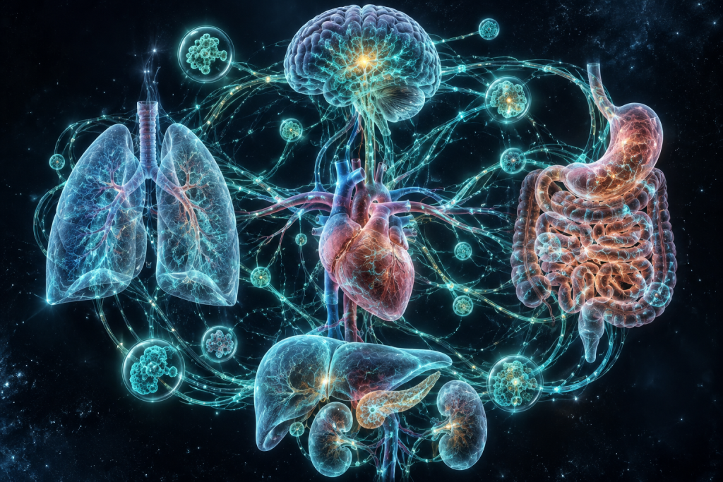
Image generated by AI
Your heart beats in perfect synchrony with your lungs, which deliver oxygen to your blood at precisely the right moment. Your digestive system knows exactly when to activate based on signals from your brain. Your kidneys adjust their filtering rates to match what your cardiovascular system demands. Yet somehow, no central conductor orchestrates this symphonic dance—your organs communicate through an intricate network of chemical messengers, electrical signals, and mechanical feedback loops that developed before you took your first breath. How does this coordination emerge from a single fertilized egg, and what happens when these finely tuned systems fail?
Understanding organ system coordination and development has never been more urgent. Developmental disorders, birth defects, and age-related diseases increasingly trace their roots to breakdowns in how our organs communicate and form. Modern medicine is beginning to leverage this knowledge to regenerate damaged tissues, engineer artificial organs, and even reverse certain developmental abnormalities. With advances in genetic sequencing, organoid technology, and computational modeling, scientists are decoding the rules that govern how complex biological systems organize themselves—knowledge that could transform medicine and help us build functioning replacements for failed organs.
What Is Organ System Coordination and Development?
Organ system coordination and development refers to the biological processes by which separate organs and tissues form during embryonic growth and then communicate to function as integrated units throughout life. During development, a cluster of identical cells must somehow “decide” to become a heart, liver, kidney, or brain—each with distinct structures and functions. Then, these differentiated organs must establish communication channels so that they can work together seamlessly. This coordination involves multiple layers of biological organization: molecular signaling pathways that activate genes at precise times, physical interactions between tissues that sculpt developing structures, and feedback mechanisms that allow mature organs to respond to one another’s changing needs. The process is deterministic enough to produce consistent human anatomy across billions of people, yet flexible enough to accommodate individual variation and environmental influences.
The conceptual foundation for understanding organ development emerged gradually through the 19th and 20th centuries. In the 1880s, German embryologist Hans Driesch performed experiments separating sea urchin cells and discovered that isolated cells could still develop into complete organisms—proving that development was governed by chemical and physical rules, not mysterious vital forces. Decades later, Hans Spemann’s work in the 1920s revealed that certain tissues, which he called “organizers,” could instruct nearby tissues to develop in specific ways through diffusible chemical signals. These experiments led to the modern understanding of embryonic induction and patterning. By the 1990s, researchers like Christiane Nüsslein-Volhard and Eric Wieschaus used genetic analysis in fruit flies to identify the molecular switches that control development, earning them the Nobel Prize and launching the modern era of developmental systems biology.
How It Works in Nature
Organ system coordination and development operate through a hierarchical cascade of molecular programs that activate with remarkable precision. In the earliest stages of embryonic development, maternal factors deposited in the egg create gradients—concentration fields of proteins that vary from one end of the embryo to the other. These gradients activate transcription factors, proteins that bind to DNA and turn genes on or off. Different concentrations of the same transcription factor can trigger different developmental outcomes, allowing the embryo to establish a coordinate system—a molecular map of where things are. As development proceeds, cells interpret these gradients and produce their own signaling molecules, creating secondary gradients that refine tissue identity and shape. This process involves dozens of conserved signaling pathways: the Wnt pathway, the Hedgehog pathway, the Notch pathway, and the BMP pathway all orchestrate different aspects of tissue formation and organ assembly.
Consider how the heart develops as a concrete example. Around the third week of human development, a small group of cells called the cardiac mesoderm receives signals indicating that it should become heart tissue. These signals activate a specific combination of transcription factors—including NKX2.5 and GATA4—that commit cells to a cardiac fate. Simultaneously, other signaling molecules organize these cardiac cells into a simple tube. Over subsequent weeks, this tube folds and subdivides, cells migrate to precise locations, and specialized regions begin to form: chambers for pumping, valves for directing flow, and specialized muscle cells for coordinated contraction. Throughout this process, cell-to-cell communication ensures that the different regions of the developing heart coordinate their growth and maturation. Electrical signals eventually develop, allowing the heart cells to fire in sequence and generate heartbeats. The entire transformation from undifferentiated cells to a fully functional organ involves thousands of genes being activated and silenced in exact temporal order—a genetically encoded choreography refined by millions of years of evolution.
Medical and Scientific Relevance
Disruptions in organ system coordination and development cause some of the most common birth defects and chronic diseases. Congenital heart disease, which affects roughly 1 percent of live births, results when developmental signaling goes awry during early cardiac formation. Neural tube defects, causing spina bifida and anencephaly, arise from failures in the coordination between neural and mesodermal tissues. Diabetes occurs partly because the pancreas fails to coordinate proper development and maintenance of its insulin-producing beta cells. Cancer itself is increasingly understood as a disease of developmental coordination gone wrong—tumor cells reactivate developmental programs that enable them to divide indefinitely, migrate through tissues, and evade immune detection. Understanding the molecular basis of these disorders has become essential to developing new treatments, and researchers are using this knowledge to create diagnostic tools that identify developmental problems before birth and interventions that might prevent or reverse them.
In current medical practice, organ system coordination knowledge enables several powerful applications. Prenatal screening for developmental abnormalities now uses 3D ultrasound and genetic analysis to detect coordination failures early in pregnancy. Regenerative medicine researchers are using developmental principles to coax stem cells into differentiating into specific cell types and assembling into functional tissue patches that could replace damaged organs. Pharmaceutical companies screen potential drugs using organoids—miniature, self-organizing versions of organs grown in petri dishes—to predict how developmental toxins might harm human development before testing in humans. Precision medicine initiatives use understanding of developmental genetics to personalize cancer treatment based on which developmental pathways a tumor has reactivated.
Recent Breakthroughs in Organ System Coordination and Development
The past three years have witnessed remarkable advances in organ system coordination research, particularly through the development of more sophisticated organoid models and the application of artificial intelligence to developmental biology. In 2022 and 2023, researchers at institutions including MIT and Cambridge successfully created human cardiac organoids that could undergo coordinated contractions and showed electrical synchronization similar to early hearts. Simultaneously, computational approaches using machine learning have begun to predict how changes in developmental genes will affect organ formation, creating a kind of “developmental atlas” that maps how different tissues coordinate their development. A landmark 2023 study used spatial transcriptomics—technology that reveals which genes are active in specific locations within developing tissues—to create high-resolution maps of how the human heart coordinates tissue interactions during early development. These advances reveal an unexpectedly flexible and self-organizing process, where organs can partially compensate for genetic variations, suggesting that developmental robustness involves multiple redundant communication channels.
Current research frontiers are pushing toward even more ambitious goals. Scientists are working to understand how organ systems communicate across the entire embryo—how development of the heart influences kidney formation, or how the developing nervous system coordinates with other organ systems to ensure they form in proper relationship to one another. Researchers are also investigating why developmental coordination becomes less precise with maternal age or in response to environmental toxins, which might explain why some people are more susceptible to developmental diseases. Another open question involves understanding how developmental programs persist in adult tissues: how do mature organs continue to communicate and maintain homeostasis using mechanisms similar to those that built them during development? These questions promise to unlock new therapeutic strategies for both congenital and acquired organ diseases.
Why Organ System Coordination and Development Matters for the Future
As our population ages and organ failure becomes an increasingly common cause of death and disability, understanding organ system coordination becomes essential to solving the transplant crisis and extending healthy human lifespan. Currently, over 900,000 people await organ transplants in the United States alone, yet only about 40,000 transplants occur annually. If researchers can harness developmental principles to grow replacement organs from patients’ own cells—organs that would be immunologically compatible and ethically uncontroversial—they could transform medicine. Beyond transplantation, understanding how organs develop and coordinate offers insights into aging itself: many age-related diseases involve gradual loss of coordination between organ systems, as the molecular communication channels that developed during embryogenesis accumulate damage and dysfunction. By understanding how coordination is established and maintained, we might identify intervention points to preserve or restore these connections in aging tissues.
However, significant challenges remain before these therapeutic possibilities become clinical reality. Growing complex organs in the laboratory is vastly more difficult than growing simple tissues—a liver or kidney has intricate internal structure with multiple distinct cell types precisely organized in space, and current organoid technology still cannot fully recapitulate this complexity. The genetic and environmental factors that can derail developmental coordination are numerous and often interact in complex ways, making it difficult to predict which individuals will suffer developmental problems. Ethical questions also loom: as we develop the ability to edit developmental genes or grow replacement organs, we must grapple with questions about which interventions are appropriate, who has access to them, and how to prevent misuse of this powerful technology.
Key Takeaways
- Organ system coordination and development is the process by which separate organs form during embryonic growth and establish chemical and electrical communication networks to function as integrated units throughout life.
- Development operates through hierarchical cascades of molecular signaling pathways, where concentration gradients of proteins provide positional information that guides tissue formation and organ assembly.
- The most promising application is regenerative medicine and organ engineering—using developmental principles to grow replacement organs from stem cells that could address the critical shortage of donor organs.
- Recent breakthroughs include sophisticated organoid models showing coordinated function, computational approaches that predict developmental outcomes, and spatial mapping techniques that reveal precisely how tissues coordinate during development.
- Understanding organ system coordination is essential for addressing birth defects, age-related diseases, and could enable the creation of replacement organs, potentially transforming medicine for millions of people with organ failure.
Explore TED Talks on Organ System Coordination and Development:
TED content is used under CC BY-NC-ND 4.0. © TED Conferences, LLC.
Frequently Asked Questions
How do organs communicate with each other during development if there is no central conductor directing the process?
Organs communicate through three primary mechanisms: chemical messengers (hormones and signaling molecules), electrical signals (particularly in the nervous system), and mechanical feedback loops that respond to physical forces. These signals are produced by developing tissues and detected by neighboring organs, creating a self-organizing system that coordinates growth and function without centralized control.
What molecular signals guide organ systems to develop in the correct sequence and timing during fetal development?
Developmental signaling molecules such as morphogens, growth factors, and transcription factors diffuse through developing tissues and activate specific genes at precise times and locations. These chemical gradients and signaling cascades create a temporal and spatial program that determines when each organ system forms and how it coordinates with others.
Why do birth defects and developmental disorders often involve multiple organ systems rather than just one?
Because organ systems share common developmental signaling pathways and depend on coordinated communication during formation, a disruption in one shared signal can affect multiple organs simultaneously. For example, a genetic mutation affecting a growth factor receptor may impact heart, kidney, and lung development all at once, since these systems rely on overlapping molecular mechanisms.
How are organoids and computational models helping scientists understand organ system coordination?
Organoids are miniature, three-dimensional tissue structures grown from stem cells that recapitulate early developmental interactions, allowing researchers to observe how different cell types communicate and organize in controlled conditions. Computational models integrate data from these experiments with genetic and molecular information to simulate complex developmental processes and predict how disruptions in signaling pathways lead to coordination failures.