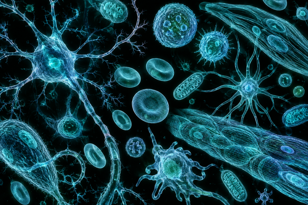
Image generated by AI
Your body contains roughly 37 trillion cells, yet fewer than 200 fundamentally different types. A neuron in your brain looks nothing like a muscle cell in your heart, despite both arising from the same genetic blueprint. How does this radical transformation happen, and how do scientists now map this cellular landscape with precision? The answers lie in understanding cellular diversity and the revolutionary techniques that allow us to identify and classify the trillions of distinct cell types that make up complex organisms.
For decades, biologists classified cells using crude methods—peering through microscopes or measuring basic chemical markers. Today, we’re entering an era where we can identify individual cell types by analyzing thousands of genes simultaneously, revealing a far more intricate cellular world than anyone imagined. This explosion of knowledge is reshaping medicine, from cancer treatment to neurodegenerative disease research, and it’s happening faster than most people realize.
What Is Cellular Diversity and Cell Type Identification?
Cellular diversity refers to the remarkable variety of specialized cell types that emerge from a single organism’s genome. Every cell in your body contains the same DNA, yet a pancreatic beta cell that produces insulin looks and behaves radically differently from a retinal photoreceptor that detects light. This specialization occurs through differential gene expression—the same genes are turned on and off in different combinations, creating distinct cellular identities. Cell type identification is the process of systematically categorizing these diverse cells based on their molecular signatures, morphology, and function. It’s detective work at the molecular level, where scientists determine what makes a cell uniquely itself.
The modern era of cellular classification began in earnest in the 1960s when scientists developed techniques to culture and examine individual cell types in isolation. Early pioneers like Paul Ehrlich and Santiago Ramón y Cajal used staining methods to distinguish neurons from supporting cells in the brain. However, the real revolution arrived in the 1990s and 2000s with the development of flow cytometry, which allowed researchers to sort cells based on fluorescently labeled markers. These tools were crude by today’s standards, but they opened a door: if we could identify cells by their surface proteins, perhaps we could create a comprehensive cellular atlas of entire organisms.
How It Works in Nature
The diversity of cell types emerges through a process called differentiation, where cells become increasingly specialized as an organism develops. During embryonic development, stem cells—which are relatively unspecialized—receive chemical signals from their environment that instruct them to adopt specific identities. These signals activate specific transcription factors, proteins that bind to DNA and switch particular genes on while suppressing others. Over time, a cascade of molecular events locks in a cell’s identity, progressively restricting its potential. A cell destined to become a neuron activates genes for neurotransmitters and ion channels while silencing genes for muscle contraction, creating a stable, self-reinforcing state that persists even after the original signals fade.
Think of differentiation like a series of increasingly narrow doorways. A stem cell can walk through any door, but once it enters a specific hallway marked “neuron,” certain molecular locks close behind it, making other pathways inaccessible. Each choice narrows future options. A neural stem cell can still become either a neuron or a glial cell, but once committed to becoming a neuron, it’s highly unlikely to transform into a pancreatic cell. The molecular signatures that mark this commitment—patterns of gene expression, surface proteins, and metabolic preferences—are what scientists now use to identify cell types with precision.
Medical and Scientific Relevance
Understanding cellular diversity has profound implications for medicine. Diseases often result from the loss, corruption, or malfunction of specific cell types. Cancer, for instance, frequently arises when a cell type loses the molecular constraints that normally prevent uncontrolled division. Diabetes occurs when insulin-producing beta cells fail or die. Parkinson’s disease involves the degeneration of dopamine-producing neurons in specific brain regions. By precisely identifying which cell types are affected and understanding their normal molecular signatures, researchers can develop targeted therapies that restore function or prevent damage. Additionally, cell type identification reveals previously unknown subtypes within apparently homogeneous populations, showing that a “liver cell” is actually dozens of distinct types, each with different vulnerabilities and therapeutic targets.
Modern cell type identification is crucial for regenerative medicine and drug development. Researchers creating tissues from stem cells must verify that differentiation is proceeding correctly, generating the right proportions of the right cell types. In cancer research, identifying the specific cell of origin for a tumor—is it a beta cell? An immune cell? A supporting fibroblast?—determines treatment strategy. Pharmaceutical companies now use cell type identification to understand which cells their drugs affect, helping predict side effects and efficacy. The immune therapy revolution, where personalized cancer treatments train a patient’s own immune cells to attack tumors, depends on identifying and characterizing the specific immune cell populations in each individual’s blood and tissue.
Recent Breakthroughs in Cellular Diversity and Cell Type Identification
The past three years have witnessed a transformative acceleration in our ability to categorize cellular diversity. The most consequential development is the maturation of single-cell RNA sequencing (scRNA-seq), a technology that measures gene expression in individual cells rather than in averaged tissue samples. Early versions generated thousands of data points per cell; current methods capture tens of thousands of genes per cell, providing an extraordinarily detailed molecular profile. This has revealed that what biologists once considered a single cell type often contains multiple subtypes with distinct molecular signatures and likely distinct functions. For example, researchers studying the human brain have identified hundreds of distinct neuron and glial subtypes, many previously unrecognized. Simultaneously, spatial transcriptomics—techniques that measure gene expression while preserving information about where cells sit in tissue—have shown that a cell’s location and neighborhood profoundly influence its identity and behavior.
The Human Cell Atlas, an international collaborative effort launched around 2016, represents an audacious attempt to create a comprehensive map of all human cell types and their locations in different tissues and organs. Major advances in 2023 and 2024 include detailed atlases of the immune system, parts of the nervous system, and developing embryos. Researchers are also increasingly using machine learning and artificial intelligence to automatically classify cells based on their molecular signatures, moving beyond hand-curation toward scalable, standardized approaches. Open questions driving current research include: How many true cell types exist in the human body? Do cell types represent discrete categories or a continuum? How stable are cell type identities, and under what conditions do cells change type?
Why Cellular Diversity and Cell Type Identification Matters for the Future
The implications of comprehensive cellular cataloging extend far beyond immediate medical applications. As we build increasingly detailed maps of cellular diversity, we gain insight into how complex organs function as integrated systems. The brain isn’t just neurons; it’s a carefully balanced ecosystem of dozens of neuronal types and supporting glial cells, each playing specific roles in processing information and maintaining neural health. Understanding this diversity at the molecular level could reveal vulnerabilities targeted by neurodegenerative diseases or explain why some brains are more resilient than others. In evolutionary terms, cellular diversity itself is a fascinating frontier—comparing cellular landscapes across species can reveal which cell types are ancient and conserved versus recently evolved, providing clues about how animal body plans diversified.
Challenges remain formidable. Current methods are expensive and require specialized equipment, limiting their use to well-funded research centers. Identifying cell types relies on molecular signatures that may not fully capture functional identity—two cells with identical gene expression profiles might behave differently in the living body. Standardizing cell type definitions across laboratories and species remains an ongoing challenge; one researcher’s “beta cell subtype 1” might be another’s “subtly distinct variant.” Perhaps most fundamentally, we must move beyond classification toward understanding causation: identifying that a particular cell type exists is only the first step. Determining what that cell type does, why it’s necessary, and how to maintain or restore it in disease requires integration with functional studies, animal models, and human research.
Key Takeaways
- Cellular diversity—the existence of roughly 200 fundamentally different cell types in the human body—emerges through differential gene expression, where the same genome creates radically different cellular identities through selective activation and silencing of genes.
- Cell differentiation works through a cascade of molecular decisions guided by environmental signals and transcription factors that progressively restrict cellular potential, locking cells into specialized states.
- The most promising application is personalized medicine: precisely identifying which cell types are affected in disease and developing targeted therapies that restore function rather than causing widespread side effects.
- Single-cell RNA sequencing and spatial transcriptomics have revolutionized our ability to catalog cellular diversity over the past five years, revealing far more subtlety in cell type organization than previously recognized, with international efforts like the Human Cell Atlas systematically mapping all human cell types.
- Comprehensive understanding of cellular diversity will reshape medicine from cancer to neurodegeneration, guide regenerative therapy development, and reveal fundamental principles of how complex multicellular life organizes itself at the molecular level.
Explore TED Talks on Cellular Diversity and Cell Type Identification:
TED content is used under CC BY-NC-ND 4.0. © TED Conferences, LLC.
Frequently Asked Questions
How can cells with identical DNA become so structurally and functionally different?
Cellular specialization occurs through differential gene expression, where different genes are selectively turned on or off in different cell types through epigenetic mechanisms and transcription factor regulation. This allows the same genetic blueprint to produce a pancreatic beta cell or retinal photoreceptor despite identical underlying DNA sequences.
What modern techniques allow scientists to identify individual cell types with greater precision than traditional microscopy?
Contemporary methods like single-cell RNA sequencing (scRNA-seq) and multi-omics analysis enable researchers to examine thousands of genes simultaneously in individual cells, revealing their molecular signatures far more accurately than older light microscopy or basic chemical marker approaches. This technology has uncovered that cellular diversity is far more complex than previously appreciated.
Why is mapping cellular diversity important for medical research and treatment?
Understanding the specific characteristics of different cell types enables researchers to develop more targeted therapies for diseases like cancer and neurodegenerative disorders by identifying which cells are abnormal and how to selectively treat them. Precise cell type identification also reveals disease mechanisms at the cellular level that were previously invisible to researchers.
How many distinct cell types do humans actually have, and how did scientists determine this number?
Humans have fewer than 200 fundamentally different cell types despite containing approximately 37 trillion individual cells, determined through systematic classification using genetic, morphological, and functional characteristics. This classification continues to be refined as new sequencing technologies reveal previously unidentified cell subtypes within broader categories.